

Two-Day programme brings together experts, faculty members and research scholars to explore the capabilities of high-end equipment facilities at the Institution of Eminence Interdisciplinary Research Centre (IoE-IDRC) University of Hyderabad, leading to cutting edge interdisciplinary research.
The Institution of Eminence Interdisciplinary Research Centre (IoE-IDRC) organised a two-day series of expert technical and interactive sessions on 3rd and 4th September 2026, on the advanced high-end equipment facilities established under the Institution of Eminence. The sessions held in the IDRC auditorium brought together experts from the original equipment manufacturers, faculty members, research scholars and students of the University of Hyderabad to explore the capabilities of sophisticated facilities available for cutting-edge and interdisciplinary research.
Prof. M. Ghanashyam Krishna Director, IoE in his welcome address emphasized that the facilities were unique to the University system in the country and encouraged all stakeholders to use the facilities for cutting edge research problems that would enhance the research profile and impact of the University of Hyderabad as an Institution of Eminence
Day One: Advanced electron microscopy and x-ray diffraction facilities
The first day, held on 3rd September 2026, focused on the facilities for focused ion beam-SEM), Analytical Transmission electron microscopy (A-TEM) and micro- x-ray diffractometer (micro-XRD).
Prof. J.P. Gautam of the School of Engineering Sciences and Technology (Faculty in-charge of the facilities and Chairperson of the technical committee) explained briefly the challenges in establishing the facilities, the capabilities and the process of accessing them.
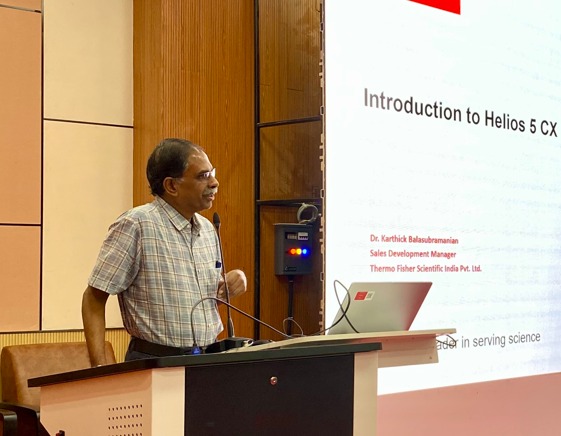
Dr. Karthick Balasubramanian representing Thermofisher Scientific, India made the presentation on the FIB-SEM (Model Helios 5 CX Dual Beam FIB-SEM with EDS), Mr. Dinesh Adire of JEOL, India made the presentation on the ATEM (model JEOL NEOARM200F Aberration-Corrected Analytical Transmission Electron Microscope (TEM)) and Dr. T. Pravin representing M/s IR Technologies the Indian partner for M/s Rigaku Japan presented the capabilities of the micro-XRD (Model SmartLab μHR Micro-XRD with CMF optics).
Dr. Venkata Girish Kotnur (co-Faculty in charge of the facilities) of the School of Engineering Sciences and Technology moderated the session.
The Analytical TEM enables researchers to examine materials at near atomic scales and obtain detailed information about their structure, micro/ nano structure and composition. It is particularly useful for advanced studies in nanotechnology, materials science and engineering, life sciences, physics, chemistry and related fields.
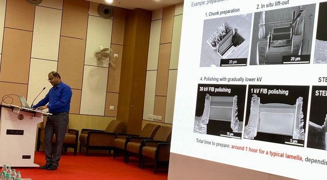
The Dual Beam FIB-SEM combines imaging and precision material processing capabilities, allowing researchers to study both surface and internal features of samples. Its analytical capabilities also support elemental characterisation and advanced materials research. The equipment is specifically useful for preparing samples for the Analytical TEM facility
The Micro-XRD enables X-ray diffraction studies from very small regions of a sample, making it useful for understanding local structural and crystalline properties of materials.
Following the presentations, there was an active and vibrant interactive session between the 50 participants and the experts.
Day Two: Advanced Optical Imaging and spectroscopy facilities
The second day, held on 4th September 2026, focused on advanced optical imaging and spectroscopy platforms.
Prof. Akash Gulyani of the School of Life Sciences (faculty in charge of the facilities and Chairperson of the technical committee) explained briefly the challenges in setting up the facilities, the capabilities, the process of accessing them and moderated the session. It was also mentioned that integrating all the components is underway and will be completed very shortly.
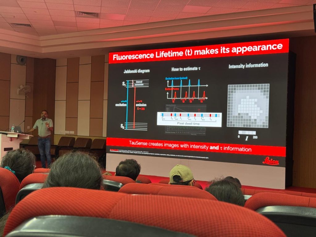
The session was conducted by Dr. Manoj Manna, Leica Microsystems, India. The session introduced participants to advanced imaging solutions based on the Leica STELLARIS platform, including two-photon microscopy, confocal imaging, spectral imaging, live cell imaging as well as fluorescence lifetime (FLIM) imaging (FALCON system). This Leica Stellaris-based system is capable of multi-photon imaging through Stellaris DIVE platform as it is coupled to an ultrafast femtosecond laser. It is equipped with a pulsed supercontinuum ‘white light’ laser source for seamless FLIM, FLIM-FRET and spectral scanning. MICA is a widefield and confocal imaging system that is capable of high throughput high content imaging of live and fixed cells and tissues in a sample protecting, incubating environment.
Following the presentations, there was an active and vibrant interactive session between the 80 participants and the experts.
The two-day programme provided a valuable platform for researchers to familiarise themselves with advanced scientific technologies and understand their potential applications across diverse disciplines. The interactive discussions enabled participants to engage directly with technical specialists, exchange ideas and explore how the sophisticated platforms can support ongoing and future research at the University.
The programme also highlighted the importance of interdisciplinary approaches in modern research, where advanced microscopy, imaging and analytical techniques are increasingly contributing to discoveries across physics, chemistry, engineering, life sciences, nanotechnology and earth sciences.
The two-day sessions concluded with a renewed emphasis on collaboration, knowledge exchange and greater utilisation of advanced research facilities, further contributing to the University’s vision of fostering high-quality and interdisciplinary research.